I-Wall brings congenital abnormalities in the unborn child vividly to life for parents
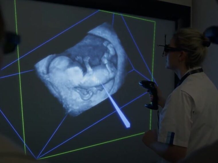
With the new virtual reality screen at the Obstetrics and Gynaecology outpatient clinic at Erasmus MC, it’s as if parents step into the womb. The I-Wall magnifies the secrets of the unborn fetus as a hologram. It is the centerpiece of the world’s first outpatient center for innovative prenatal imaging.
Every year, thousands of expectant mothers with concerns about their child’s health come to the Fetal Medicine outpatient clinic at Erasmus MC. Thanks to the new I-Wall, known as the system with the 3-meter wide and 2-meter high virtual reality wall, every detail of the developing child is clearly visible. It’s as if you can touch the child.
In the waiting room, expectant parents sit anxiously waiting. An ultrasound has just been taken of their little one at thirteen weeks. The midwife had a feeling something wasn’t quite right. Now, a team of healthcare providers is examining the images with special equipment. “We collectively determine whether there are congenital abnormalities and what this means for the child and for the expectant parents,” say gynecologists Annemarie Mulders and Melek Rousian.
World’s First
Until now, healthcare providers mainly assessed the unborn child using gray 2D images. Thanks to developments in ultrasound techniques, Mulders and Rousian, along with colleagues, have been researching ways to improve imaging in early pregnancy in recent years. And with success: the 3D ultrasound technique with virtual reality on the large screen is now being introduced in the outpatient clinic. “A world’s first,” says fetal medicine physician Carsten Pietersma, who hopes to graduate with this research this year.
Shortly after, the parents and healthcare providers stand together in a dark room in front of a sort of movie screen. With special 3D glasses, a gigantic hologram appears in the room, measuring a square meter. In reality, the unborn child is only 6.6 centimeters long. But everything is there. “Unlike during an ultrasound, the baby is still on the screen because it’s a recording of the earlier ultrasound. The baby’s movements do not obstruct the study of the images on the I-Wall,” says fetal medicine physician and doctoral candidate Kristel Zandbergen.
Alarm Bell
Zandbergen skillfully rotates the hologram on the screen using the computer’s special joystick. “We assess the baby from head to toe,” she says. With the pointer, she scrolls through the organs to detect any congenital abnormalities. She looks for possible growth differences and easily measures the volume of the bladder. “It’s like measuring the contents of a balloon. I perform these measurements after the ultrasound has been taken. This way, I can take time to calmly view the images without it being a burden for the pregnant woman.”
“The position of a foot or an extra finger is usually better seen in 3D”
From heart defects to clubfoot. “If we find a congenital abnormality in the first trimester of pregnancy, it can be serious. But not necessarily,” says Pietersma. “Occasionally, we can reassure parents.” The position of a foot or an extra finger is usually better seen in 3D than in 2D and does not seem to have major consequences at first glance. “But,” adds Rousian, “it is an alarm bell for us: Something is wrong. Investigate further.”
Expectation
Mulders explains that a DNA condition cannot be seen on the outside. But its manifestations sometimes can be. “Think, for example, of fluid around the child or in the neck. Through a puncture in the mother’s abdomen, that can be investigated on the same day. Tissue from the placenta or amniotic fluid is then extracted from the uterine cavity using a needle.”
The doctors ask themselves every time: what can we expect? With multiple abnormalities, a genetic condition is more often involved, Rousian knows. “With an extra finger, you can live to be a hundred. And an opening in the abdominal wall can be surgically closed. But there are also abnormalities that will significantly decrease the quality of life. The severity of the anomaly, whether caused by a genetic condition or not, generally determines the prognosis for the child.”
Considered Choice
Early in pregnancy, looking ensures that parents know what’s going on in a timely manner. They can prepare themselves and make informed choices. Many emotions come with congenital abnormalities. “Guilt, questions, and doubts. We hope to better support parents with the images,” says Mulders.
“We hope to better support parents with the images”
If it turns out that a child cannot live independently outside its mother’s womb, then the parents decide whether to continue the pregnancy. “Seeing their child as a hologram and giving it a name might even help some parents in coping,” concludes Zandbergen.
Healthy
After viewing the images, the healthcare providers take the parents to the adjacent consultation room to discuss things calmly. The couple breathes a sigh of relief because their baby is healthy. The suspicion of a twisted foot on the 2D ultrasound turns out not to be a clubfoot, as the I-Wall showed.
About the I-Wall
The I-Wall at the Fetal Medicine outpatient clinic is the result of years of research at Erasmus MC. In 2005, researchers used the I-Space, a room with 3D technology on three walls and the floor. Bioinformatician Anton Koning of Erasmus MC then developed the V-Scope application specifically to better analyze 3D scans, such as ultrasounds. This VR technology has been used since 2012 on a small scale on a 3D TV at the Obstetrics and Fetal Medicine outpatient clinic. The Generation R research cohort also uses an I-Wall.
Date: January 15, 2025
Source: Amazing Erasmus MC


